How Serial Sleep Studies Reveal Your Narcolepsy’s Hidden Timeline Through Sleep Study Changes Reflecting Narcolepsy Progression
Story-at-a-Glance
- Narcolepsy type 1 shows remarkable stability in testing, with 88% diagnostic accuracy and 80% reliability in serial sleep studies—far more consistent than type 2
- Brain wave disruptions in delta and theta activity distinguish narcolepsy patients from healthy controls during NREM sleep
- Progressive cortical thinning appears in multiple brain regions, particularly the prefrontal cortex and hippocampus, with patterns varying by age of onset
- Pediatric patients display unique markers: increased wake time after sleep onset, more frequent awakenings, and additional stage shifts compared to adults
- Daytime continuous polysomnography offers a promising alternative to traditional MSLT testing, especially valuable for children
- Predictable progression patterns emerge through serial monitoring, opening doors to personalized treatment timing
Main Body
Picture this: An 11-year-old girl experiences daytime sleepiness and moderate snoring at night. She receives an initial diagnosis of narcolepsy that wasn’t supported by proper testing protocols. Her family seeks a second opinion at the University of Florida. Repeat sleep studies conducted under appropriate conditions revealed something crucial—the initial diagnosis was incorrect. Understanding sleep study changes reflecting narcolepsy progression proved essential for accurate diagnosis, demonstrating why serial monitoring matters so profoundly.
The landscape of narcolepsy diagnosis is undergoing a fundamental shift. Dr. Emmanuel Mignot at Stanford won the 2023 Breakthrough Prize for discovering that narcolepsy stems from the loss of approximately 70,000 hypocretin-producing neurons in the hypothalamus. He and other leading researchers are championing a new paradigm. Rather than viewing narcolepsy as a static condition captured in a single diagnostic snapshot, we’re learning to read its evolving signature through serial sleep studies—what experts call the disorder’s “hidden timeline.”
Save This Article for Later – Get the PDF Now

The Dynamic Nature of Sleep Architecture in Narcolepsy
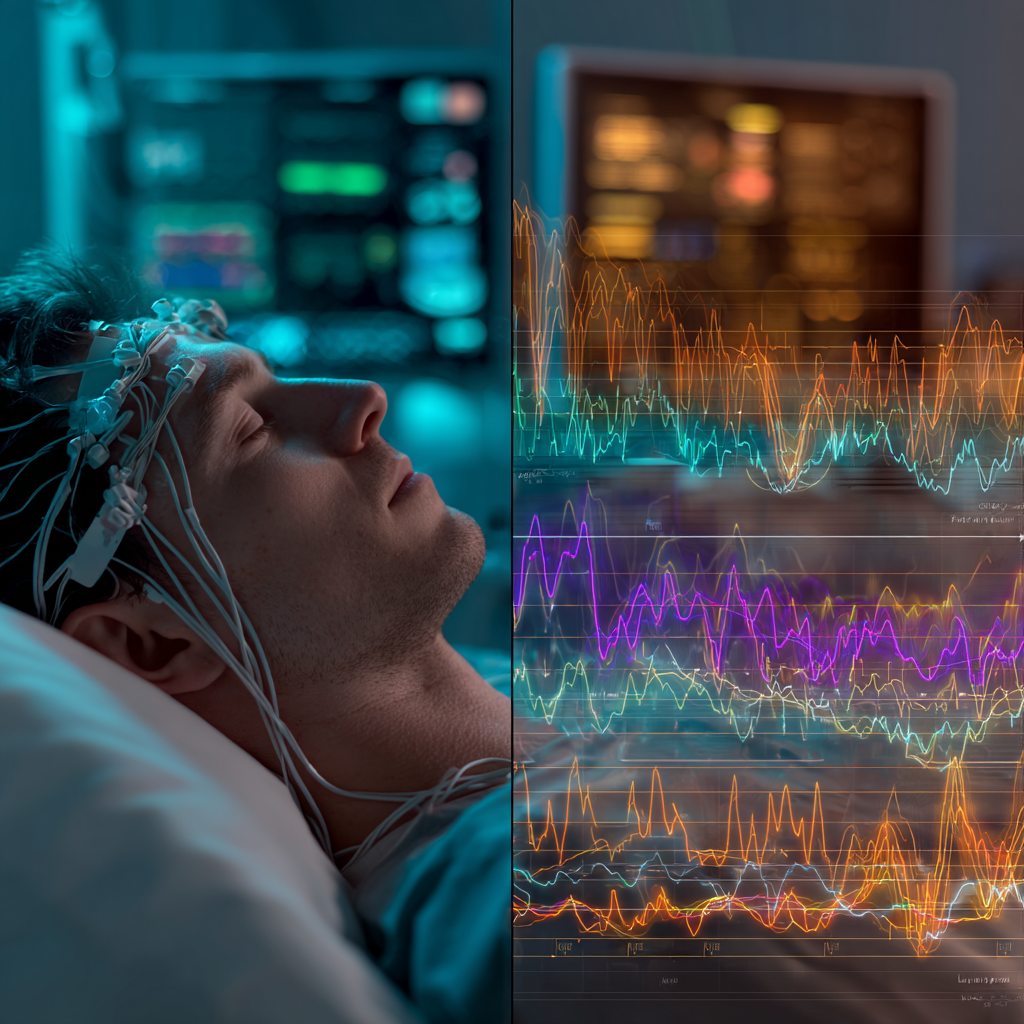
Fascinating patterns emerge when we examine polysomnographic data longitudinally. Research from Seoul National University revealed something striking: delta activity in control groups decreased consistently from the first to fourth NREM episodes throughout the night. Narcolepsy patients, however, showed this decrease only from the first to third episodes. This suggests a fundamental disruption in sleep homeostasis—not just academic minutiae, but a window into how sleep study changes reflecting narcolepsy progression manifest at the cellular level.
Theta waves tell an equally compelling story. Healthy individuals show theta activity that increases progressively through all four NREM periods. In narcolepsy patients, this increase halts after the third period. Think of it as watching a symphony where certain instruments gradually fall silent. The overall performance continues, but something essential is missing.
What makes these findings particularly intriguing? Their potential link to hypocretin deficiency. High amplitude theta wave bursts might directly relate to the loss of hypocretin neurons—one of the major pathophysiological features of narcolepsy. This connection between microscopic neuronal loss and macroscopic sleep wave patterns represents a crucial bridge in understanding disease progression.
Age-Dependent Progression Patterns: Not All Narcolepsy Is Created Equal
The story takes an unexpected turn here. Longitudinal brain imaging studies demonstrate that early-onset cases show different progression patterns compared to late-onset cases. Participants exhibited widespread progressive cortical thinning in multiple brain regions including the prefrontal cortex. This discovery challenges our one-size-fits-all approach to monitoring the condition.
Pediatric populations present unique considerations. A comprehensive meta-analysis of 108 studies revealed something important: children and adolescent narcolepsy patients demonstrate increased wake time after sleep onset (WASO), awakening numbers (AWN), and stage shifts (SS) compared to adults. These aren’t merely developmental differences—they’re distinct markers of how sleep study changes reflecting narcolepsy progression manifest across the lifespan.
Dr. Birgitte Rahbek Kornum from the University of Copenhagen leads a team that discovered autoreactive CD8 T cells in narcolepsy patients’ blood. Their work provided crucial proof of the disorder’s autoimmune nature. She emphasizes that understanding these age-related patterns could revolutionize early intervention strategies. For patients seeking ways to work with their condition, behavioral therapy approaches that target sleep architecture in narcolepsy offer complementary strategies alongside medical monitoring.

The Reliability Question: When Sleep Studies Tell Different Stories
Now, let’s address the elephant in the sleep lab: test reliability. Research from Danish and Stanford sleep centers found that narcolepsy type 1 showed excellent test-retest reliability. Both MSLTs were positive for narcolepsy in 78% of unmedicated NT1 patients. Narcolepsy type 2 patients, however, showed poor reliability—only 18% consistency.
This disparity has profound implications for clinical practice.
Consider the frustration of patients with NT2 who might receive different diagnoses from repeated testing. NT2 cases changed diagnostic categories to idiopathic hypersomnia 26% of the time. They shifted to negative MSLT results 57% of the time upon retesting. This isn’t a failure of the test itself but rather a reflection of the heterogeneous nature of non-hypocretin-deficient hypersomnolence disorders.
The implications extend beyond individual diagnosis. Insurance companies often require specific test results for coverage approval. Yet these findings suggest that a single negative test in suspected NT2 shouldn’t close the diagnostic door. Serial monitoring becomes not just helpful but essential for capturing the true nature of the disorder through sleep study changes reflecting narcolepsy progression.
Revolutionary Approaches: Beyond Traditional Testing
Innovation in sleep medicine is accelerating rapidly. Italian researchers at the University of Bologna validated daytime continuous polysomnography (D-PSG) as an alternative to traditional MSLT. This proves particularly valuable for pediatric patients where obtaining reliable MSLT results can be challenging. Imagine the relief for parents who no longer need to subject their young children to multiple overnight stays and daytime napping sessions.
Additionally, Stanford researchers developed neural network analysis capable of automated sleep stage scoring with 87% accuracy—better than any individual human scorer. The system also identifies narcolepsy markers based on unusual sleep stage overlaps with 91% sensitivity and 96% specificity. This isn’t just about efficiency; it’s about catching subtle patterns that human eyes might miss.
Clinical Pearls from Serial Monitoring
Through analyzing thousands of serial sleep studies, several key patterns have emerged that clinicians should monitor:
Early-stage indicators:
- Shortened REM latency (≤15 minutes) on nighttime polysomnography—occurs in about one-third of NT1 patients with high specificity for narcolepsy diagnosis
- Increased sleep fragmentation even before cataplexy onset
- Subtle changes in sleep efficiency that precede daytime symptoms
Progression markers:
- Progressive increase in N1 sleep percentage
- Decreased slow wave sleep percentage and reduced cyclic alternating pattern rate
- Evolution of REM sleep intrusions from isolated incidents to frequent occurrences
Treatment response indicators:
Serial studies can reveal whether interventions are genuinely improving sleep architecture or merely masking symptoms. This distinction proves crucial when adjusting medication regimens or considering newer treatments like Lumryz. The FDA-approved once-nightly sodium oxybate formulation eliminates the need for middle-of-the-night dosing.
The Immunological Timeline: What Triggers Progression?
The autoimmune hypothesis of narcolepsy has evolved from speculation to established fact. Yet questions remain about progression triggers. The discovery of autoreactive T cells in some healthy individuals suggests something fascinating: autoimmunity lies dormant in many of us but requires specific triggers for activation. This finding fundamentally changes how we think about disease surveillance.
Environmental triggers appear to play a crucial role. The well-documented increase in narcolepsy cases following the 2009 H1N1 pandemic and certain vaccinations provides a natural experiment in disease triggering. Understanding how these triggers translate to progressive changes in sleep architecture—the very sleep study changes reflecting narcolepsy progression that we’ve been discussing—could help identify at-risk individuals before symptom onset.

FAQ
Q: What exactly are sleep study changes reflecting narcolepsy progression?
A: These are measurable alterations in brain wave patterns, sleep stage transitions, and REM sleep timing that evolve as narcolepsy develops and progresses. Unlike a static snapshot, serial sleep studies reveal how the disorder changes sleep architecture over months and years.
Q: How often should someone with suspected or confirmed narcolepsy undergo sleep studies?
A: Based on research showing different reliability rates between NT1 and NT2, experts suggest annual monitoring for NT2 patients. Stable NT1 patients may test every 2-3 years, with more frequent monitoring during treatment changes.
Q: What’s the difference between polysomnography and MSLT in tracking progression?
A: Polysomnography records overnight sleep patterns including brain waves, breathing, and movement—revealing structural sleep changes. The MSLT measures daytime sleep tendency and REM sleep timing across multiple naps, showing functional impairment. Both provide complementary information about disease progression.
Q: Can serial sleep studies predict who will develop cataplexy?
A: While not definitively predictive, certain patterns may indicate higher risk. Early REM sleep onset (within 15 minutes) during nighttime polysomnography and specific EEG spectral changes in delta and theta activity may signal increased cataplexy risk.
Q: What is D-PSG and how does it compare to traditional MSLT?
A: Daytime continuous polysomnography (D-PSG) is an alternative diagnostic tool that continuously monitors sleep patterns during the day rather than using scheduled naps like MSLT. Research shows promise especially for pediatric patients where traditional MSLT can be challenging.
Q: Are the brain changes seen in narcolepsy reversible?
A: Current evidence suggests that cortical thinning and hippocampal changes are progressive and likely irreversible. This emphasizes the importance of early diagnosis and treatment to potentially slow progression.
Q: How do sleep study changes differ between children and adults with narcolepsy?
A: Pediatric patients typically show more sleep fragmentation with increased wake time after sleep onset and more frequent stage shifts compared to adults. This possibly reflects the developing nervous system’s response to hypocretin loss.