Serotonin Dysregulation in Narcolepsy Sleep Architecture: Why Your Brain’s Chemical Messenger Can’t Keep You Awake
Story-at-a-Glance
• Narcolepsy stems from the loss of hypocretin neurons, which normally activate serotonin-producing cells—creating a cascading chemical imbalance that affects both wakefulness and muscle control • Serotonin receptor availability increases dramatically during sleep in narcolepsy patients, opposite to what happens in healthy individuals, suggesting the brain compensates for chronically low serotonin activity during wakefulness • The link between serotonin and narcolepsy explains why antidepressants that boost serotonin levels are the primary treatment for cataplexy—they’re replacing a missing chemical signal • Recent breakthrough research reveals narcolepsy patients lose not only hypocretin neurons but also norepinephrine-producing cells in the brainstem, compounding the serotonin dysfunction • Understanding serotonin’s role offers hope for new orexin receptor agonist medications currently in clinical trials, which may address the root cause rather than just managing symptoms
In 2017, researchers at Kanazawa University made a startling discovery using genetically modified mice. When they artificially increased serotonin release in a specific brain region—the amygdala—cataplexy attacks almost completely vanished. This wasn’t just about dampening emotions or reducing muscle weakness. The serotonin was doing something far more sophisticated: quieting the very brain circuits that trigger sudden collapse when people with narcolepsy experience joy.
It’s a finding that fundamentally changed how we understand serotonin dysregulation in narcolepsy sleep architecture. The neurotransmitter we typically associate with mood and well-being plays a critical, previously underappreciated role in keeping narcolepsy’s most dramatic symptom at bay.
The Hypocretin-Serotonin Connection: A Missing Link in Your Brain’s Wakefulness System
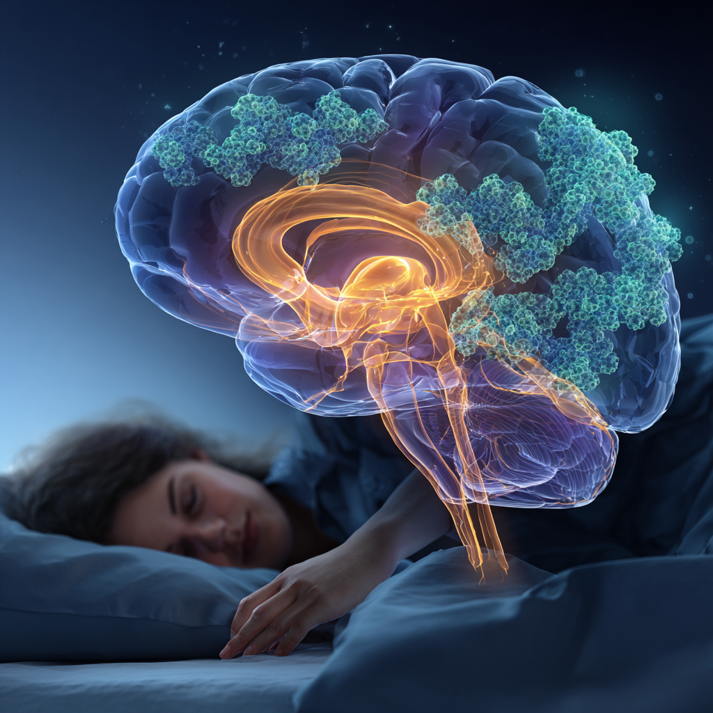
To understand how serotonin becomes dysregulated in narcolepsy, we first need to understand what’s lost. Dr. Emmanuel Mignot at Stanford University—who won the 2023 Breakthrough Prize in Life Sciences for his work—discovered that narcolepsy stems from the loss of roughly 70,000 specialized neurons in the hypothalamus. These neurons produce hypocretin (also called orexin), a wake-promoting chemical that serves as the brain’s master switch for staying alert.
Here’s where serotonin enters the picture. Those hypocretin neurons don’t work in isolation—they actively stimulate other brain regions that produce neurotransmitters essential for wakefulness, including serotonin, norepinephrine, and dopamine. When hypocretin neurons die off (likely through an autoimmune process), the serotonin system loses a critical activating signal.
The result? Serotonin-producing neurons in the dorsal raphe nucleus become inconsistent. During waking hours, serotonin levels may be lower than normal. The brain struggles to maintain the neurotransmitter balance needed for sustained alertness, contributing to the excessive daytime sleepiness that defines narcolepsy.
When Your Brain Chemistry Runs Backward: Increased Serotonin Receptors During Sleep
A fascinating 2006 study using PET scans revealed something unexpected about serotonin in narcolepsy patients. Researchers at the Epilepsy Research Centre in Australia measured serotonin receptor availability in patients during both wakefulness and sleep. What they found contradicted normal patterns.
In healthy individuals, serotonin release promotes wakefulness and suppresses REM sleep. But in narcolepsy patients, serotonin receptor availability increased significantly during sleep compared to wakefulness—across the whole brain and in specific regions like the temporal cortex and cingulate cortex.
Think of it this way: when serotonin is actively released and binding to receptors, those receptors become “occupied” and appear less available on brain scans. The increased receptor availability during sleep suggests that serotonin levels drop even further when narcolepsy patients try to rest. It’s as though the brain’s serotonin system is running in reverse, unable to maintain the normal oscillation between sleep and wake states.
This finding helps explain why people with narcolepsy experience such fragmented sleep at night. The serotonin dysregulation in narcolepsy sleep architecture doesn’t just affect daytime wakefulness—it disrupts nighttime sleep quality too, creating a vicious cycle of poor rest and excessive sleepiness.

The Amygdala Connection: How Serotonin Stops Emotions from Triggering Collapse
Perhaps the most elegant demonstration of serotonin’s role in narcolepsy comes from understanding cataplexy—those sudden episodes of muscle weakness triggered by strong emotions like laughter or excitement.
During REM sleep, our muscles are normally paralyzed by circuits in the lower brainstem and spinal cord. This prevents us from acting out our dreams. In healthy people, norepinephrine and serotonin block these paralysis circuits during wakefulness. But when hypocretin neurons are lost, levels of both neurotransmitters drop. This allows the paralysis circuits to activate inappropriately—even when someone is fully awake and experiencing a moment of joy.
The breakthrough research from Kanazawa University showed that serotonin neurons don’t directly control muscle tone. Instead, they regulate activity in the amygdala, the brain’s emotional processing center. When someone with narcolepsy sees something funny or experiences excitement, their amygdala responds excessively. It’s missing the dampening effect of serotonin input from hypocretin neurons.
This excessive amygdala activity then triggers the brainstem circuits that cause muscle weakness. It’s a domino effect: loss of hypocretin → reduced serotonin signaling → overactive amygdala → inappropriate activation of REM sleep paralysis circuits → cataplexy.
Understanding this pathway explains why antidepressants that increase serotonin levels are effective treatments for cataplexy. Particularly selective serotonin reuptake inhibitors (SSRIs) and serotonin-norepinephrine reuptake inhibitors (SNRIs) work by compensating for the missing hypocretin signal through boosting serotonin availability.
The 1976 Study That Predicted What We Know Today
Sometimes science moves in circles. Back in 1976—decades before hypocretin was even discovered—researchers examined serotonin metabolism in narcolepsy patients. They found that serotonin metabolism was “considerably disturbed,” with increased serotonin concentration in platelets and cerebrospinal fluid, but decreased excretion of 5-hydroxyindoleacetic acid (5-HIAA, serotonin’s main metabolite) in urine.
At the time, this seemed like a curious anomaly. We now understand it as evidence of compensatory changes—the body trying to maintain adequate serotonin levels despite the loss of hypocretin’s activating influence. The brain may produce more serotonin or hold onto it longer, but without hypocretin’s coordinating signal, it can’t deploy that serotonin effectively across the sleep-wake cycle.
These early metabolic studies complement modern imaging research, painting a picture of a serotonin system that’s not simply “low” or “high,” but rather dysregulated—unable to oscillate properly between sleep and wake states.
The Double Loss: When Norepinephrine Cells Disappear Too
If you thought the story of serotonin dysregulation in narcolepsy sleep architecture was complete, 2025 brought a significant plot twist. Dr. Jerome Siegel at UCLA, who first reported hypocretin loss in human narcolepsy in 2000, announced another groundbreaking discovery. All narcolepsy patients also lose norepinephrine-producing neurons in the locus coeruleus, a brainstem region critical for alertness and muscle tone.
This 46% decrease in norepinephrine neurons isn’t caused by hypocretin loss—it appears to be a separate target of the same autoimmune process. The finding is crucial because norepinephrine and serotonin work together to block the muscle paralysis circuits during wakefulness. Losing both systems creates a perfect storm for cataplexy.
Interestingly, this norepinephrine loss doesn’t occur in genetically narcoleptic mice or dogs—only in human patients. It suggests that human narcolepsy involves a more complex autoimmune attack than we previously understood, one that may require addressing both forebrain and brainstem pathologies for optimal treatment.
The clinical implications are significant. Current narcolepsy treatments focus heavily on boosting catecholamine (dopamine and norepinephrine) activity through medications like amphetamines or modafinil. While these help with alertness, they may not fully address the serotonergic dysfunction that contributes to both fragmented sleep and cataplexy.
Save This Article for Later – Get the PDF Now

Real Patient Experiences: When Medications Modify Serotonin
Clinical studies provide glimpses into how modulating serotonin affects real patients. A 2010 Stanford study of childhood narcolepsy tracked 53 patients and their treatment outcomes. While stimulants like modafinil helped with sleepiness, serotonin-active medications showed the most dramatic effects on cataplexy.
Venlafaxine, an SNRI that increases both serotonin and norepinephrine, had a 68% continuation rate among children who tried it—one of the highest for any medication. Traditional SSRIs like fluoxetine showed more modest results, with many patients trying them but fewer continuing long-term.
This real-world data aligns with our understanding of serotonin dysregulation in narcolepsy sleep architecture. Medications that boost serotonin help, but those that address both serotonin and norepinephrine systems (like SNRIs) often work better—likely because they’re compensating for both neurotransmitter deficiencies simultaneously.
I should note here that individual responses vary considerably. Some patients report that SSRIs trigger rebound cataplexy when stopped suddenly, a phenomenon called status cataplecticus. Others find that these medications lose effectiveness over time or cause unwanted side effects like sexual dysfunction or emotional blunting.
This variability probably reflects differences in how severely each patient’s serotonin system is affected—and reminds us that serotonin dysregulation in narcolepsy isn’t uniform across all patients.
The Promise of Orexin Agonists: Fixing the Root Problem
The most exciting development in narcolepsy treatment doesn’t aim to boost serotonin directly. Instead, researchers are developing medications that activate orexin receptors—essentially replacing the missing hypocretin signal.
In July 2025, Takeda announced overwhelmingly positive Phase 3 results for oveporexton (TAK-861), the first orexin receptor 2 agonist to show broad efficacy. The medication met all primary and secondary endpoints, improving wakefulness, cataplexy, and quality of life. Unlike current symptom-management approaches, oveporexton addresses the core orexin deficiency that drives narcolepsy.
Here’s what makes this particularly relevant to serotonin: by activating orexin receptors, these medications should restore the normal stimulation of serotonin neurons. In theory, this would re-establish proper serotonin oscillation between sleep and wake, rather than just artificially boosting serotonin levels with antidepressants.
Other companies are pursuing similar approaches. Eisai’s E2086 and Harmony Biosciences’ TPM-1116 are both in clinical development. Early data shows these medications can improve daytime wakefulness with tolerable side effect profiles, though we’re still learning about their full effects on sleep architecture and REM sleep dissociation.
The question that keeps me up at night (ironically): will restoring hypocretin signaling be enough to reverse the norepinephrine neuron loss that Dr. Siegel’s team discovered? Or will patients need combination therapies that address both the missing hypocretin signal and the depleted norepinephrine/serotonin systems?
The Psychiatric Comorbidity Connection
There’s another dimension to serotonin dysregulation in narcolepsy sleep architecture that deserves attention: the high rates of depression and anxiety in narcolepsy patients.
A 2025 review found that 26-28% of narcolepsy patients have comorbid depression, significantly higher than the general population. While some of this stems from the challenges of living with a chronic sleep disorder, the neurobiology suggests a deeper connection.
Hypocretin neurons normally excite serotonergic neurons. When hypocretin is deficient, serotonin levels drop—and serotonin plays a crucial role in mood regulation. The same neurotransmitter deficiency that causes excessive sleepiness and cataplexy may contribute directly to depressive symptoms.
This creates a clinical dilemma. SSRIs prescribed for depression in narcolepsy patients may help both mood and cataplexy—a rare win-win in medicine. However, doctors must monitor carefully, as some patients experience paradoxical effects or worsening of certain symptoms.
The relationship between hypocretin deficiency and emotional regulation also extends to the amygdala’s heightened reactivity. Narcolepsy patients’ amygdalas respond excessively to emotional stimuli—not just triggering cataplexy, but potentially contributing to anxiety and emotional dysregulation more broadly.
What This Means for You: Practical Implications
If you’re dealing with narcolepsy or suspect you might have it, understanding serotonin dysregulation in narcolepsy sleep architecture offers several practical insights:
First, recognize that your symptoms aren’t “just” about being tired. The neurotransmitter dysfunction affects multiple systems—wakefulness, muscle tone, mood, and sleep quality itself. This isn’t something you can power through with willpower; it’s a chemical signaling problem that needs appropriate treatment.
Second, medications that affect serotonin may help with multiple symptoms simultaneously. If you’re taking an SSRI or SNRI for cataplexy, it may also improve mood and anxiety—or vice versa. However, these medications don’t work for everyone, and finding the right combination often requires patience and close work with a knowledgeable sleep medicine specialist. Understanding how narcolepsy medications work can help you make more informed treatment decisions.
Third, be aware that treatments affect the serotonin system differently. Stimulants like modafinil primarily boost dopamine and may indirectly affect serotonin. Sodium oxybate works through the GABA system but has downstream effects on multiple neurotransmitters. The new orexin agonists should restore more normal serotonin function by replacing the missing hypocretin signal. Understanding these mechanisms can help you have more informed conversations with your healthcare provider.
Fourth, consider that sleep quality matters as much as daytime alertness. The serotonin dysregulation doesn’t just make you sleepy during the day—it fragments your sleep at night. Improving nighttime sleep architecture through behavioral interventions, optimal medication timing, and good sleep hygiene practices can have ripple effects on daytime function.
Looking Forward: The Future of Narcolepsy Treatment
We’re living through a remarkable moment in narcolepsy research. The discoveries about hypocretin loss, serotonin dysregulation, and now norepinephrine neuron loss are painting an increasingly complete picture of what goes wrong in narcolepsy—and how we might fix it.
The FDA approval of sodium oxybate for pediatric patients in October 2024 marked progress in addressing narcolepsy across the lifespan. The pending approval of orexin agonists like oveporexton represents a potential paradigm shift—from managing symptoms to addressing root causes.
Yet questions remain. Will orexin agonists fully normalize serotonin function? Can we prevent or reverse the autoimmune attack that causes neuron loss in the first place? How do we best treat the subset of patients with narcolepsy type 2, who have normal hypocretin levels but still experience excessive sleepiness?
These are areas where the research community continues to push forward. Dr. Mignot’s lab is investigating the specific T-cell variants involved in the autoimmune attack. Dr. Siegel’s team is working to understand how norepinephrine loss contributes to symptoms and whether it can be prevented. Researchers worldwide are exploring everything from biomarkers to new drug targets.
For those of us interested in sleep health more broadly, narcolepsy research offers profound insights. The intricate dance between hypocretin, serotonin, norepinephrine, and dopamine doesn’t only matter for narcolepsy patients—these same systems regulate everyone’s sleep-wake cycles. Understanding what happens when they fail helps us appreciate how remarkable it is that most people’s brains manage this balance so seamlessly.

FAQ Section
Q: What is serotonin dysregulation in narcolepsy sleep architecture?
A: Serotonin dysregulation in narcolepsy refers to the abnormal patterns of serotonin activity that occur when hypocretin (orexin) neurons are lost. Normally, hypocretin neurons activate serotonin-producing cells to promote wakefulness and suppress REM sleep. When these neurons die, serotonin levels become inconsistent—too low during wakefulness (contributing to sleepiness) and showing reversed patterns during sleep. This dysregulation affects both daytime alertness and nighttime sleep quality, creating the fragmented sleep-wake cycles characteristic of narcolepsy.
Q: What are hypocretin neurons and why do they matter?
A: Hypocretin neurons (also called orexin neurons) are approximately 70,000 specialized cells in the hypothalamus that produce wake-promoting peptides. They act as master regulators of the sleep-wake system, sending signals to other brain regions that produce neurotransmitters like serotonin, norepinephrine, and dopamine. In narcolepsy type 1, about 90% of these neurons are destroyed, likely through an autoimmune process, leading to the cascade of symptoms including excessive daytime sleepiness, cataplexy, and disrupted sleep.
Q: What is cataplexy?
A: Cataplexy is sudden, bilateral loss of voluntary muscle tone triggered by strong emotions—typically positive ones like laughter, excitement, or surprise. It occurs because the brain circuits that normally paralyze muscles during REM sleep become active during wakefulness. Episodes can range from subtle (slight facial drooping, slurred speech) to severe (complete collapse), but patients remain fully conscious throughout. Cataplexy is pathognomonic for narcolepsy type 1, meaning it only occurs in this specific condition.
Q: How do SSRIs help with narcolepsy symptoms?
A: Selective serotonin reuptake inhibitors (SSRIs) increase serotonin availability in the brain by preventing its reabsorption after release. In narcolepsy, this helps compensate for the reduced serotonin activity caused by hypocretin neuron loss. SSRIs are primarily used to treat cataplexy, as increased serotonin helps dampen the excessive amygdala activity that triggers muscle weakness during emotional moments. They may also help with mood symptoms common in narcolepsy patients, though not everyone responds to these medications and some experience side effects.
Q: What is the amygdala and how does it relate to narcolepsy?
A: The amygdala is a brain region crucial for processing emotions, particularly fear and pleasure. In narcolepsy, the amygdala responds excessively to emotional stimuli because it lacks the normal dampening effect of serotonin input from hypocretin neurons. This heightened amygdala activity connects to brainstem circuits that control muscle tone, triggering cataplexy when strong emotions occur. The amygdala overactivity may also contribute to anxiety and emotional dysregulation that many narcolepsy patients experience.
Q: What are orexin receptor agonists?
A: Orexin receptor agonists are a new class of medications designed to activate orexin (hypocretin) receptors in the brain, mimicking the action of the lost hypocretin neurons. Unlike current treatments that only manage symptoms, these medications address the root cause of narcolepsy type 1 by replacing the missing hypocretin signal. Drugs like oveporexton (TAK-861) selectively activate the orexin 2 receptor and have shown promising results in Phase 3 trials, improving wakefulness, reducing cataplexy, and enhancing quality of life with tolerable side effects.
Q: What is the locus coeruleus?
A: The locus coeruleus is a small brainstem region containing neurons that produce norepinephrine, a neurotransmitter crucial for alertness and muscle tone regulation. Recent research by Dr. Jerome Siegel’s team at UCLA discovered that all narcolepsy patients have approximately 46% fewer neurons in this region—a second neuron loss beyond the well-known hypocretin neuron loss. This dual loss helps explain why both alertness and muscle tone are affected in narcolepsy and suggests the autoimmune attack targets multiple brain systems.
Q: What is REM sleep and how does it relate to narcolepsy symptoms?
A: REM (rapid eye movement) sleep is the stage of sleep characterized by vivid dreams, increased brain activity, and muscle paralysis (called atonia) that prevents us from acting out our dreams. In narcolepsy, the circuits that control REM sleep become dysregulated—REM sleep can intrude into wakefulness (causing cataplexy and sleep paralysis) or occur too quickly after falling asleep (sleep-onset REM periods). This dysregulation stems from the loss of hypocretin neurons, which normally help maintain clear boundaries between wake, REM sleep, and non-REM sleep.
Q: What are 5-HT1A receptors?
A: 5-HT1A receptors are a specific type of serotonin receptor found throughout the brain, particularly in regions involved in mood and arousal. The “5-HT” designation refers to serotonin (5-hydroxytryptamine is serotonin’s chemical name), and the “1A” specifies this particular receptor subtype. In narcolepsy research, measuring 5-HT1A receptor availability using PET scans has revealed abnormal patterns—increased receptor availability during sleep suggests reduced serotonin release at that time.
Q: What are monoamine neurotransmitters?
A: Monoamine neurotransmitters are a family of chemical messengers that include serotonin, norepinephrine, and dopamine. They’re called monoamines because of their molecular structure (containing one amino group). These neurotransmitters are critical for regulating mood, alertness, movement, and the sleep-wake cycle. In narcolepsy, all three monoamine systems become dysregulated when hypocretin neurons are lost, since hypocretin normally stimulates the production and release of these neurotransmitters.
Q: What is sleep architecture?
A: Sleep architecture refers to the cyclical pattern and structure of sleep throughout the night, including the progression through different sleep stages (light sleep, deep sleep, and REM sleep) and the timing of these stages. Healthy sleep architecture involves predictable cycles lasting about 90 minutes, with increasing REM sleep as the night progresses. In narcolepsy, sleep architecture is severely disrupted—patients enter REM sleep too quickly, experience frequent nighttime awakenings, and have an unstable sleep-wake boundary that blurs throughout the 24-hour day.
Q: What is the dorsal raphe nucleus?
A: The dorsal raphe nucleus is a brainstem structure containing the majority of the brain’s serotonin-producing neurons. These neurons send projections throughout the brain and play crucial roles in regulating wakefulness, suppressing REM sleep, and modulating emotional responses. In narcolepsy, when hypocretin neurons are lost, the dorsal raphe nucleus loses a critical activating input, leading to reduced and inconsistent serotonin release that contributes to both excessive sleepiness and disrupted sleep architecture.
Q: What is an autoimmune process?
A: An autoimmune process occurs when the body’s immune system mistakenly attacks its own cells and tissues rather than foreign invaders like bacteria or viruses. In narcolepsy type 1, researchers believe that T-cells (specialized immune cells) target and destroy hypocretin neurons in the hypothalamus and norepinephrine neurons in the locus coeruleus. This attack appears to be triggered by infections (particularly influenza) in genetically susceptible individuals, especially those carrying the HLA-DQB1*06:02 genetic variant. Understanding this autoimmune mechanism opens possibilities for prevention and immune-targeted treatments.